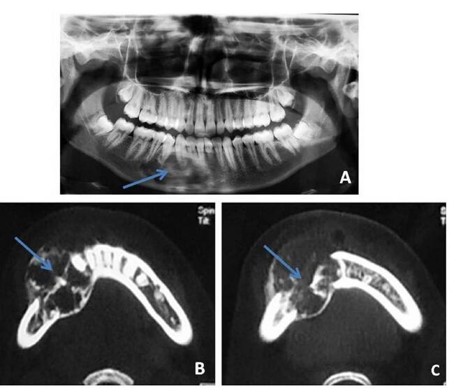
Fig. 10.

(A–C) Central giant cell granuloma: a 17-year-old lady with lower jaw swelling. Orthopantomogram (OPG) ( A ) shows multilocular, expansile radiolucent lesion (blue arrow) seen involving right side of mandible reaching the midline. Axial computed tomography (CT) images ( B, C ) at bone window settings show expansile, lytic lesion (blue arrow) seen involving right side of mandible with multiple trabeculae within the lesion.
