
Fig. 15.
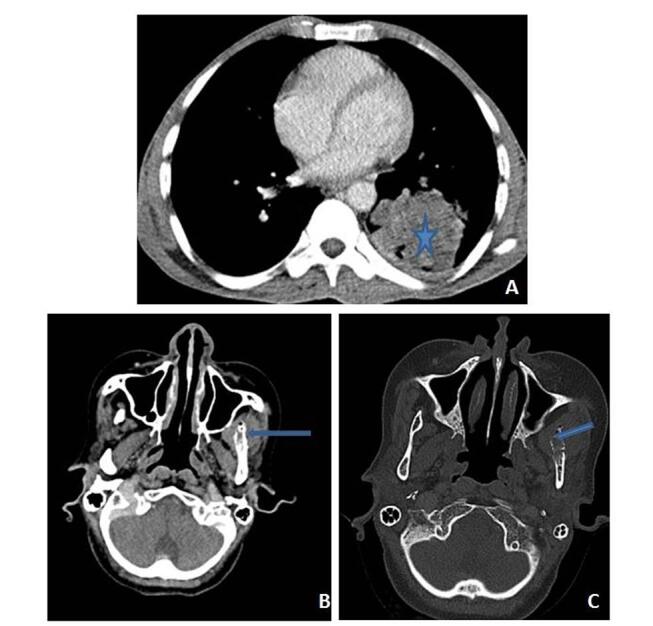
( A–C ) Metastases: a 55-year-old female patient with left lung mass. Contrast-enhanced axial ( A ) computed tomography (CT) image reveals irregularly marginated, heterogeneously enhancing mass lesion (star) in left lower lobe. Axial CT image in soft tissue window ( B ) and bone window algorithm ( C ) settings show ill-defined lytic lesion (blue arrow) with cortical destruction and associated soft tissue component involving ramus of left side of mandible.