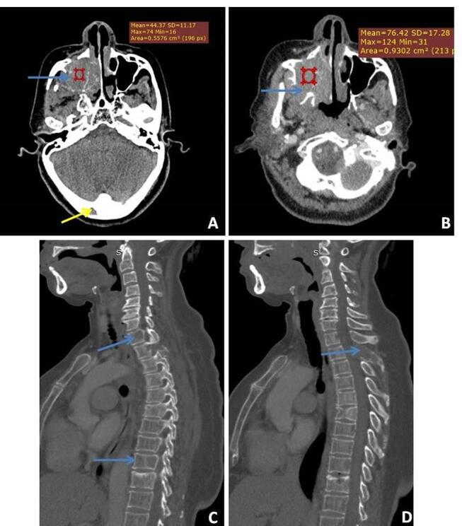
Fig. 16.

( A–D ) Multiple myeloma: a 55-year-old male with right-sided facial swelling. Axial precontrast and corresponding post contrast ( A, B ) computed tomography (CT) images show an enhancing aggressive mass involving the right maxilla with significant osseous destruction with another small lytic lesion (yellow arrow) also seen in right occipital bone. Biopsy of the larger mass revealed multiple myeloma. Evaluation of the spine ( C, D ) shows diffuse osteopenia with multiple lytic lesions involving D1, D9 vertebral bodies and posterior element of D2 vertebral body (blue arrow).
