
Fig. 3.
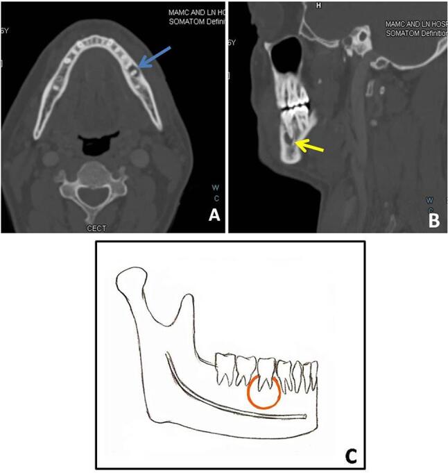
( A–C ) Radicular cyst: a 30-year-old-male with pain over the left lower jaw. Axial bone window algorithm ( A ) image shows small radiolucency around the left mandibular second molar tooth. Sagittal reformatted image ( B ) shows the periapical location of the lesion. Cyst shows a thin sclerotic rim (yellow arrow). Schematic diagram ( C ) shows the typical location of the periapical cyst (red circle) in relation to the root of the tooth. CECT, contrast-enhanced computed tomography.