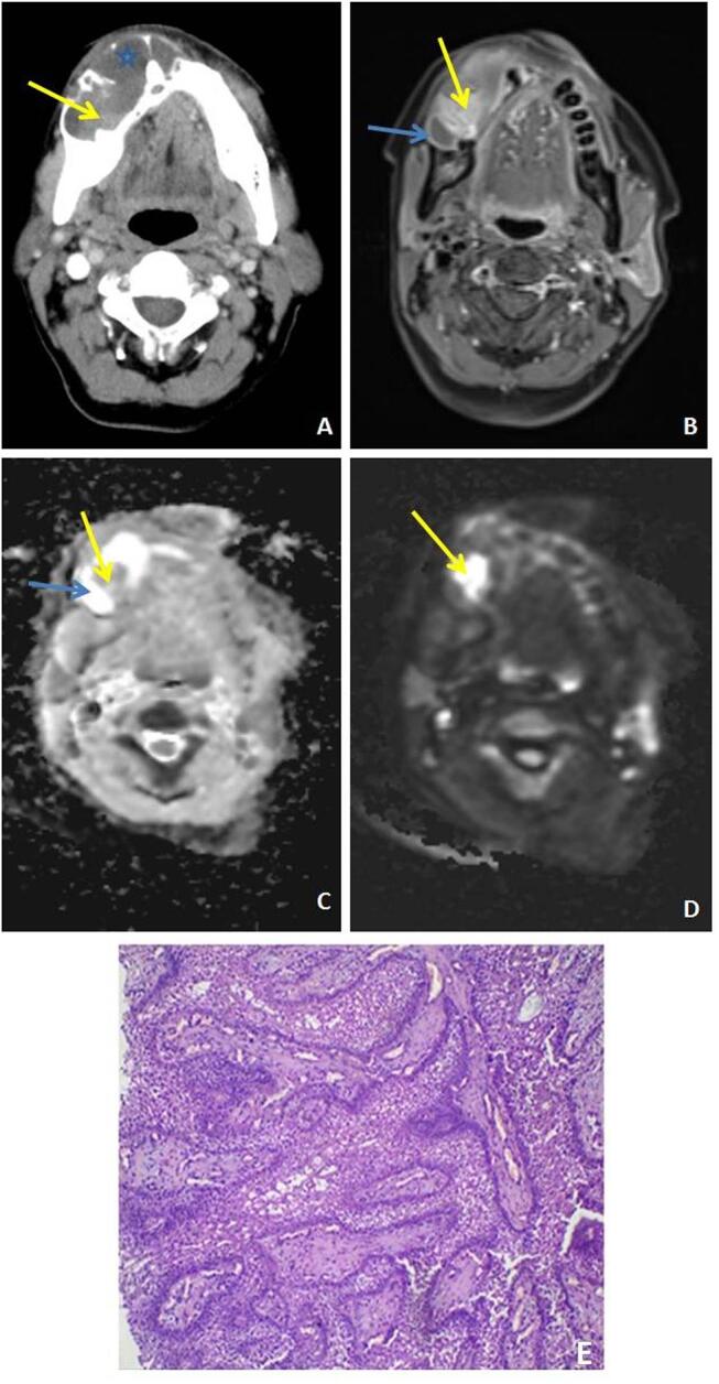
Fig. 7.

( A–E ) Ameloblastoma: axial postcontrast computed tomography (CT) ( A ) image shows a well-defined lytic lesion with predominantly buccolingual expansion. Nonenhancing cystic and enhancing solid component (yellow arrow) is seen within the lesion on CT ( A ) and postcontrast T1weighted magnetic resonance (MR) image ( B ) shows large peripherally enhancing cystic component (yellow arrow) with an enhancing solid component (blue arrow) within. The solid component of the lesion shows restricted diffusion ( C, D ). Microscopy ( E ) shows proliferating odontogenic epithelium with tall columnar peripheral cells and stellate central cells.
