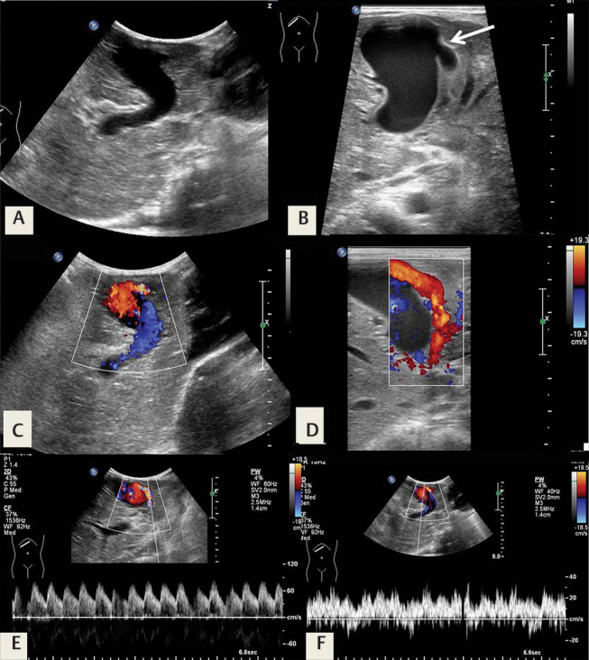
Fig. 10.

Intrahepatic arterioportal fistula in a 6-month-old infant with Down’s syndrome and abdominal distension. ( A,B ) Ultrasonography (USG) shows an aneurysmally dilated left portal vein, with an abnormal intrahepatic arterioportal communication ( arrow in B ). ( C,D ) Color Doppler shows turbulent intralesional flow. Spectral Doppler confirms ( E ) a high-velocity, low-resistance flow in the feeding artery and ( F ) chaotic, arterialized flow in the dilated vein.
