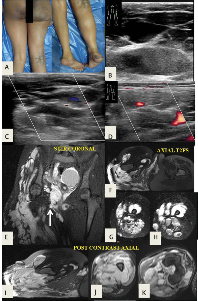
Fig. 22.

A 14-year-old girl with Klippel–Trenaunay syndrome. ( A ) The right lower limb is enlarged, with port wine stains and varicose veins. ( B–D ) Ultrasonographic (USG) Doppler shows multiple dilated vascular channels, with slow flow. ( E ) Short tau inversion recovery (STIR) coronal and ( F–H ) fat-suppressed axial T2-weighted magnetic resonance (MR) images demonstrate soft-tissue hypertrophy and extensive venous malformations involving the limb muscles, with right gluteal and intrapelvic component ( arrow in E ). These lesions are T2 hyperintense and ( I–K ) show enhancement on delayed postcontrast fat-suppressed T1 axial images.
