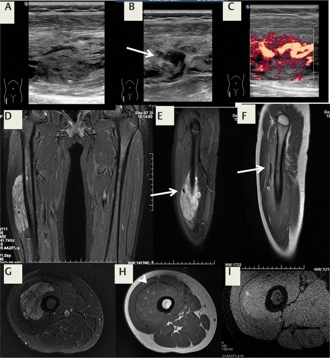
Fig. 23.

Suspected fibroadipose vascular anomaly (FAVA) in an 11-year-old girl with pain. ( A ) Ultrasonography (USG) reveals a heterogeneous intramuscular lesion with ( B ) a dilated intralesional vein and ( C ) venous channels. ( D ) Magnetic resonance imaging (MRI) short tau inversion recovery (STIR) coronal image shows the hyperintense lesion; the dilated vein is seen on ( E ) sagittal STIR and ( F ) T1 images. The lesion shows mild hyperintensity on axial ( G ) T2-weighted fat-suppressed image, ( H ) areas of hyperintensity on T1, and ( I ) mild contrast enhancement. The patient is awaiting surgical resection.
