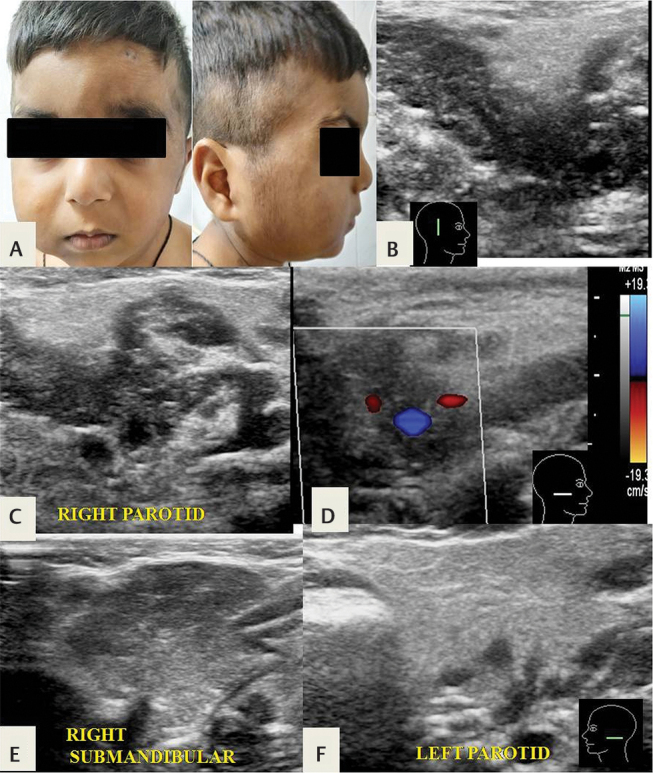
Fig. 24.

( A ) Plexiform neurofibroma presenting as a facial swelling in a 5-year-old boy with family history of neurofibromatosis type I. ( B,C ) Ultrasonography (USG) shows a lobulated, serpiginous, trans-spatial hypoechoic lesion partly within and partly surrounding the right parotid gland, ( D ) without significant intralesional vascularity. ( E ) The normal right submandibular and ( F ) left parotid glands are shown for comparison.
