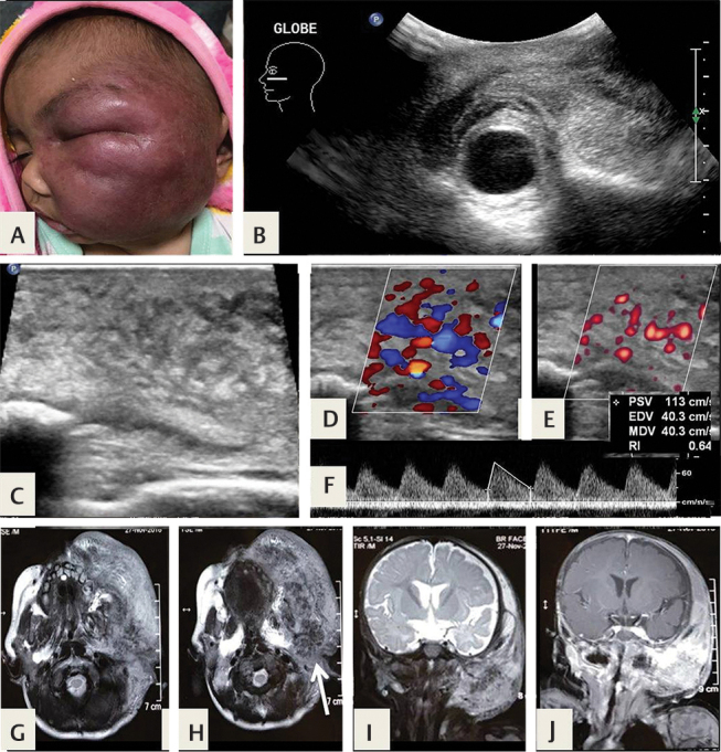
Fig. 6.

( A ) A large, infiltrative facial hemangioma in a 6-month-old infant, which developed soon after birth and grew to involve the entire left face. ( B,C ) Ultrasonography (USG) shows an ( D,F ) infiltrative, hyperechoic high-flow lesion. ( G,H ) Axial and ( I ) coronal T2-weighted (T2W) magnetic resonance (MR) images confirm an infiltrative lesion with multiple flow voids ( arrow ), involving the left parotid gland, buccal, and masticator spaces, ( J ) with intense enhancement.
