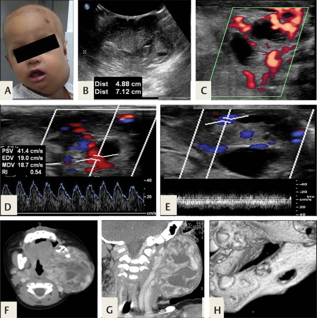
Fig. 8.

Biopsy-proven solitary fibrous tumor/ hemangiopericytoma ( A ) A 4-month-old infant presented with a rapidly growing swelling of recent onset, not typical of hemangioma. ( B,C ) Ultrasonography (USG) shows a large soft-tissue lesion with cystic areas, internal vascularity, ( D,E ) both arterial and venous flow. ( F,G ) Contrast-enhanced computed tomography (CECT) confirms a vascular soft-tissue lesion with ( H ) associated lytic lesion in left hemimandible, suggesting a locally aggressive tumor.
