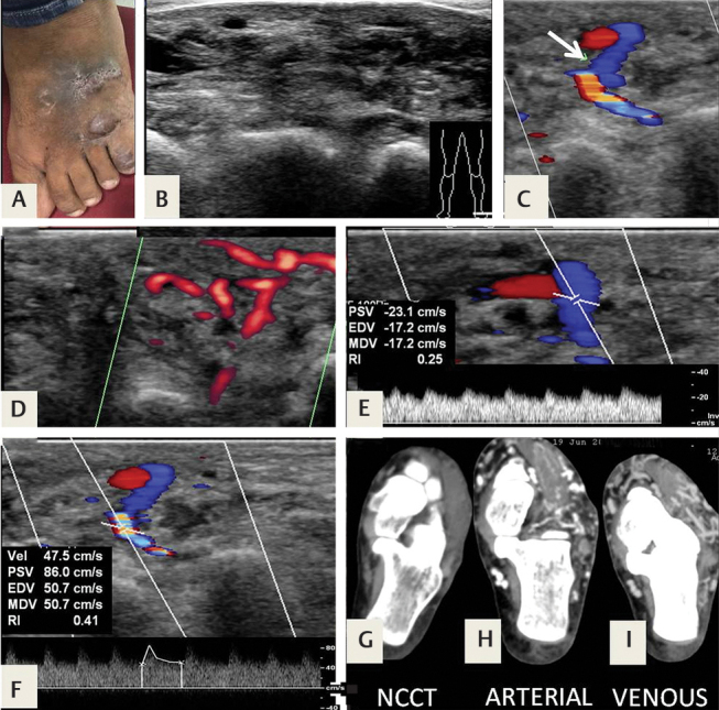
Fig. 9.

( A ) An arteriovenous malformation (AVM) in an 18-year-old man, who presented with a vascular swelling over his foot. ( B–D ) Ultrasonography (USG) showed multiple vascular channels with both arterial and venous flow. ( E ) There was arterialization of venous flow, whereas the arteries showed low-resistance flow with ( F ) a resistive index (Rl) of 0.41. ( G–I ) Computed tomography (CT) angiography showed multiple enlarged vessels, and early filling of venous channels in the arterial phase.
