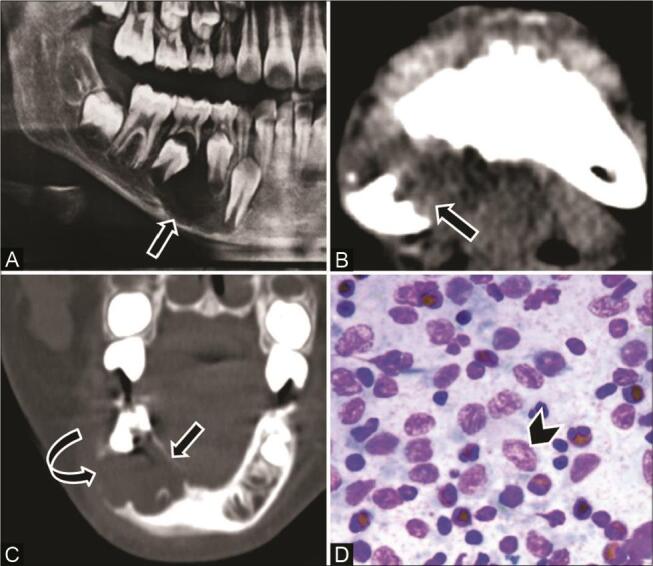
Fig. 10.

Langerhans cell histiocytosis in a 9-year-old boy with a painful swelling right lower jaw, and clinical diagnosis of dentoalveolar abscess. ( A ) Orthopantomograph (OPG) shows a well-defined radiolucent lesion ( arrow ) in the right hemimandible adjacent to unerupted premolars. ( B ) Computed tomography (CT) of the axial soft-tissue window and ( C ) coronal multiplanar reconstruction (MPR) bone window show a single well-marginated scooped-out lytic lesion ( arrow ) in the right posterior mandible with areas of cortical thinning ( curved arrow ), with no periosteal reaction or associated soft tissue. ( D ) Cytopathology reveals Langerhans cells ( arrowhead ) in the background of the eosinophils and lymphocytes.
