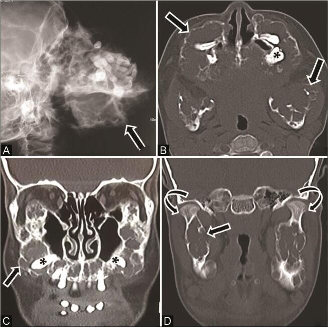
Fig. 15.

Cherubism in an 11-year-old girl with painless progressive swelling of the jaw since 2 years of age. ( A ) Skull lateral radiograph shows gross expansile bone remodeling with multiple radiolucent areas in bilateral maxilla and mandible ( arrow ), causing displacement of teeth. Computed tomography (CT) of the ( B ) axial and ( C,D ) coronal multiplanar reconstruction (MPR) soft tissue and bone window show multilocular expansile lytic lesions with coarse trabecular bony pattern involving the entire maxilla, body, and ramus of mandible ( arrow ) along with displacement of teeth ( asterisk ), but preservation of the mandibular condyles ( curved arrow ).
