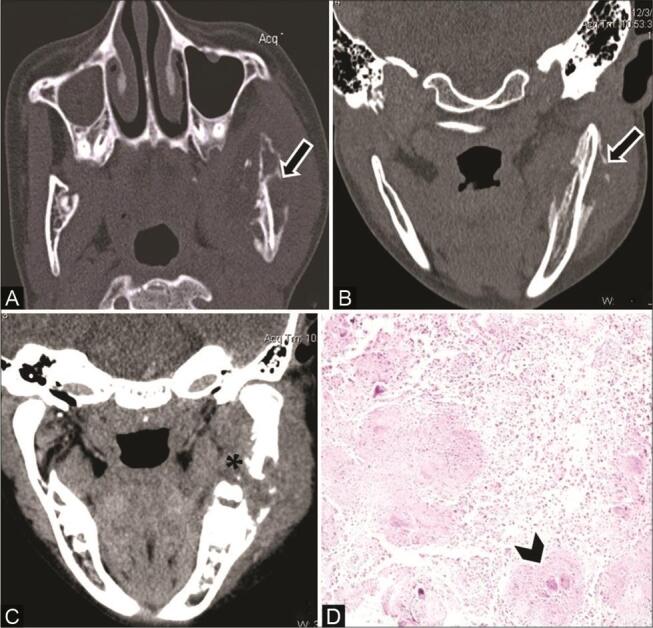
Fig. 17.

Tubercular osteomyelitis in a 12-year-old boy with diffuse hard swelling left face and positive contact history of tuberculosis (TB). ( A ) Axial computed tomography (CT) and ( B ) coronal multiplanar reconstruction (MPR) bone window show osteolytic lesions ( arrow ) causing destruction of both inner and outer cortices in the body and ramus of the left mandible with periosteal reaction. ( C ) Contrast-enhanced CT (CECT) coronal reformatted soft-tissue window reveals adjacent muscle and soft-tissue involvement with heterogeneous enhancement ( asterisk ). ( D ) Histopathological examination shows epithelioid cell granuloma with Langerhans giant cells ( arrowhead ) confirming the diagnosis of tubercular osteomyelitis.
