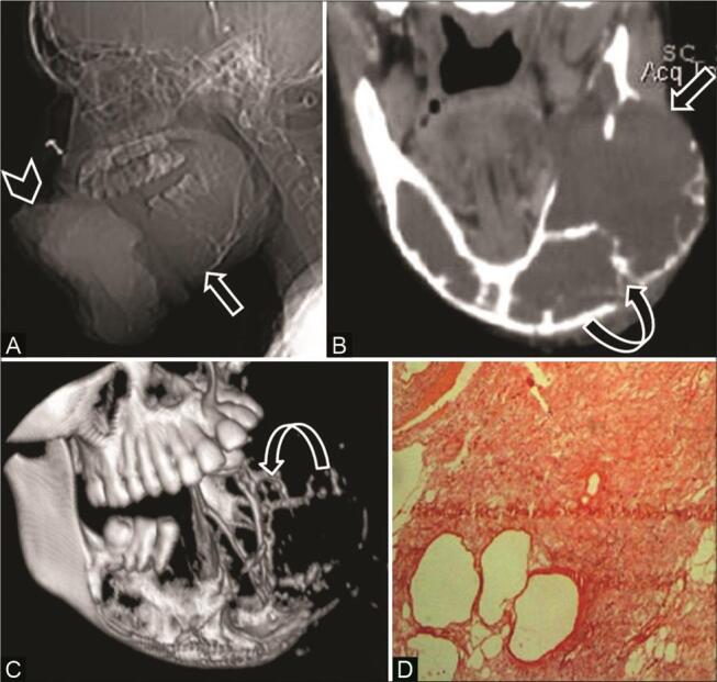
Fig. 6.

Odontogenic myxoma in a 16-year-old adolescent girl with a large painful mass lower jaw. ( A ) Computed tomography (CT) scanogram shows a large soft-tissue density mass ( arrowhead ) and expansile lytic lesion in the mandible ( arrow ). ( B ) CT coronal multiplanar reconstruction (MPR) soft-tissue window shows a large lytic expansile lesion ( arrow ) with peripheral curved bony trabeculae—”tennis racket” appearance ( curved arrow ). ( C ) 3D reformat depicts the entire tumor extent and internal bony trabeculae ( curved arrow ). ( D ) Histopathological examination reveals stellate cells in myxoid background with few thin-walled vascular spaces confirming the diagnosis.
