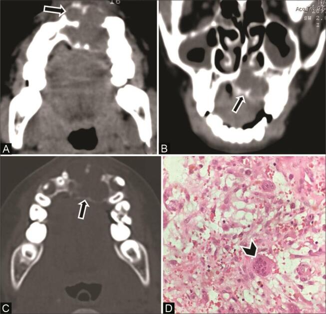
Fig. 9.

Central giant cell granuloma of the maxilla in a 12-year-old boy. ( A ) Axial computed tomography (CT) and ( B ) coronal multiplanar reconstruction (MPR) soft-tissue window show a multilocular expansile lucent lesion ( arrow ) with scalloped border predominantly in the left anterior maxilla, crossing the midline. Thin internal septations are seen with a honeycomb appearance. ( C ) CT of the axial bone window shows similar multilocular appearance ( arrow ). ( D ) Histopathological examination shows multinucleated giant cells ( arrowhead ) in fibrovascular stroma, confirming the diagnosis.
