
Fig. 1. |. Structure of the NLRP1-DPP9 complex.
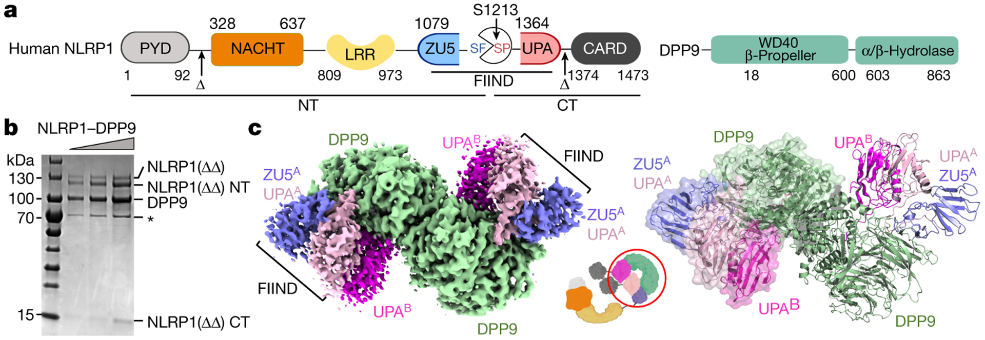
a, Domain organization. b, Representative (>3 independent experiments) SDS-PAGE of the purified NLRP1-DPP9 complex. HSP70 contamination is noted with an asterisk (*). c, Cryo-EM map (left) and the model (right) of the ternary NLRP1A-NLRP1B-DPP9 complex. The DPP9 dimer and the two copies of NLRP1 (A and B) are labelled with the colour scheme in (a). A schematic diagram (middle) denotes the entire NLRP1 and DPP9 molecules versus the ordered, resolved portions of the proteins (red circle).