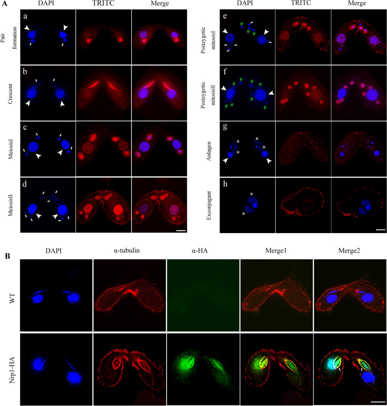
Fig. 3.
Localization of Nrp1-HA during conjugation stage. A Immunofluorescence staining of Nrp1-HA during conjugation stage. DNA was stained with DAPI. a, pair formation; b, crescent; c, meiosis I; d, meiosis II; e, postzygotic mitosis I; f, postzygotic mitosis II; g, anlagen stgage; h, exconjugant stage. B Co-localizaiton of Nrp1 and α-tubulin during early conjugation stage. The experiments were repeated three times. White arrowheads indicate parental MACs, arrows indicate MICs, stars indicate anlagen, and pounds indicate postzygotic nucleus. Scale bar, 10 µm

