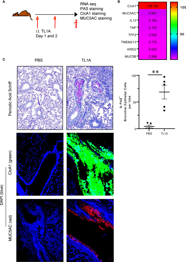
Figure 1.
TL1A induces mucus production when administered in isolation into the airways. (A) Schematic representation of the protocol used. Briefly, C57BL/6 mice were induced with 10μg of recombinant TL1A i.t. on two successive days. Mice were euthanized 24h after the last i.t. injection, and lungs were harvested for RNAseq analysis, PAS, ClcA1 and MUC5AC stains. (B) Heatmap of the top differentially regulated genes involved in mucus production, upregulated in the lungs of TL1A-induced mice. (C) Top panel: PAS stain of mucus produced in the lungs and quantified using Image Pro Premier and graphed as %PAS+ bronchial epithelial cells per lobe (right panel). Mid and Lower panels: Immunofluorescence stains of CLcA1 (green), MUC5AC (red) and nuclear DAPI (blue). All results representative of three experiments with four to six mice per group. **p < 0.005.

