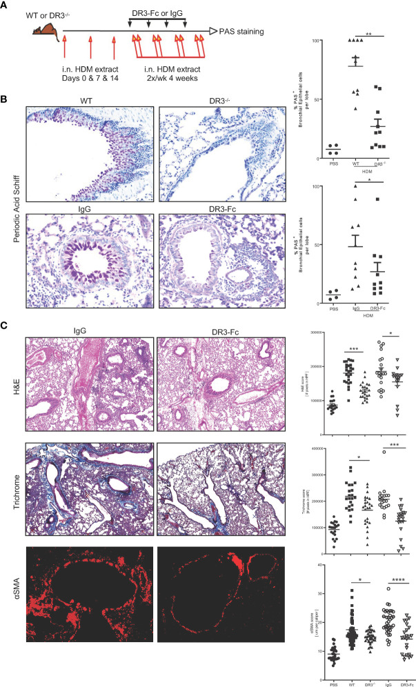
Figure 2.
Interrupting TL1A/DR3 signaling decreases mucus production in allergen-induced asthma. (A) Schematic representation of protocol used. Briefly, WT littermates and DR3-deficient mice on the C57BL/6 x 129 background were sensitized i.n. on day 0, 7, and 14 with 200 and 100 μg house dust mite extract protein in PBS, followed by chronic i.n. challenges of 50 μg of HDM protein administered twice a week for the following four weeks. Analyses were performed 24 hours after the last challenge. For neutralization of TL1A-DR3 interactions, mouse DR3-Fc or isotype control IgG were administered i.p. to WT C57BL/6 mice after the initial sensitization period starting at day 14 and were given every three days until the end of the experiment (100 μg/injection/mouse). (B) PAS stain of mucus produced in the lungs and quantified using Image Pro Premier. (C) Inflammation assessed by H&E stain (top panels), collagen deposition assessed by trichrome stain (middle panels), and smooth muscle hypertrophy assessed by αSMA immunofluorescence stain (red) (bottom panels) on lung biopsies of WT C57BL/6 mice after treatment with either IgG or DR3-Fc. Quantifications for each parameter, completed using Image Pro Premier, are shown to the right, including untreated WT C57BL/6 and DR3-/- mice (images not shown). All results representative of three experiments with five mice per group. *p < 0.05, ** < 0.005, ***p < 0.0005, ****p < 0.00005.

