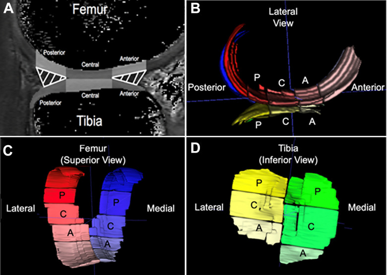
Figure 1.
(A) Weightbearing regions of interest (gray-shaded regions) were determined using the position of the meniscus in the sagittal plane: the articular cartilage overlying the anterior horn (anterior), between the anterior and posterior horns (central), and overlying the posterior horn (posterior) of the menisci. (B-D) Three-dimensional renderings of voxels determined to correspond to the segmented (B) femoral and tibial articular cartilage as seen from a lateral view in the sagittal plane, (C) femoral articular cartilage as seen from a superior view, and (D) tibial articular cartilage as seen from an inferior view. A, anterior; C, central; P, posterior.

