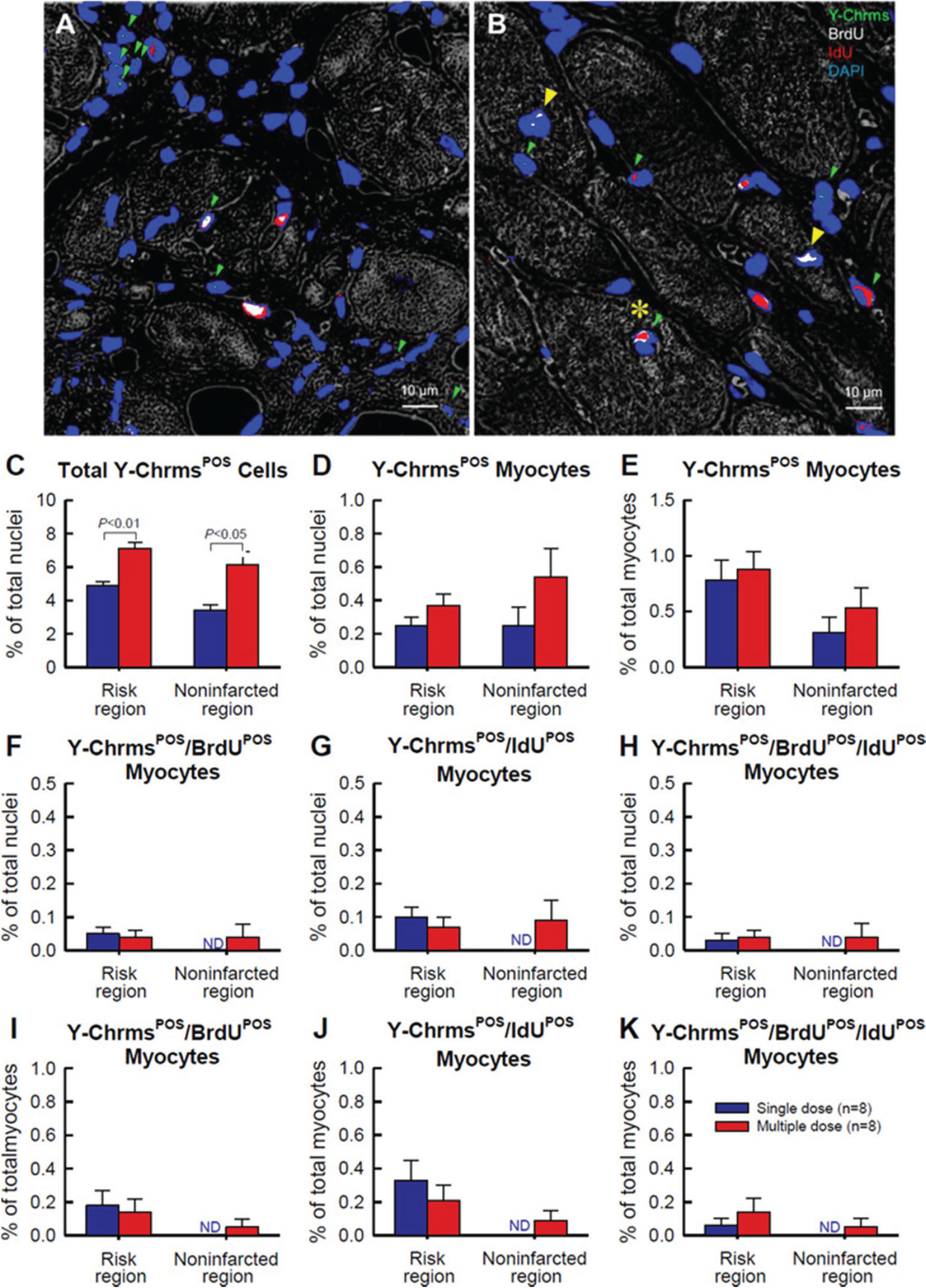
Fig. 4.

Detection of CPC retention and engraftment with fluorescence in situ hybridization (FISH) for detection of Y-chromosomes after multiple CPC administrations in rats with chronic ischemic cardiomyopathy. Male c-kit CPCs were injected into female rats with old MI 30 days after MI; three CPC injections were performed at 35-day intervals. Rats were given BrdU or IdU after CPC transplantation. (A–B) Representative confocal microscopic images acquired from the infarcted region (A) and border zone (B). Green arrowheads indicate Y-chromosome fluorescent signals (green/cyan) in nuclei. Note a cluster of five Y-chromosomePOS (Y-ChrmsPOS) nuclei in A (top left), suggesting Y-chromosomePOS cell division. Yellow arrowheads indicate BrdUPOS mature myocytes, whereas the yellow asterisk shows a Y-chromosomePOS/BrdUPOS/IdUPOS mature myocyte (B). BrdU is shown in white, IdU is shown in red, and nuclei are stained with DAPI in blue. Myocardial morphology was examined with the confocal transmitted light channel’s detector (ChD) in which the pseudocolor selected for the myocardial background in the ChD channel was gray white. (C–K) Quantitative analysis of the number of Y-chromosomePOS, BrdUPOS, and IdUPOS cells. The risk region comprises both the border zones and the infarcted region. Note that the number of newly formed (i.e., BrdUPOS and (or) IdUPOS) cardiomyocytes (regardless of their source, exogenous or endogenous) was negligible (<1% of nuclei); that is, transplantation of CPCs did not promote significant formation of new cardiomyocytes from either exogenous or endogenous (resident) cells, even after multiple CPC doses. Data are means ± SEM. Bar is 10 μm. Reproduced with permission from Tokita et al. (2016).
