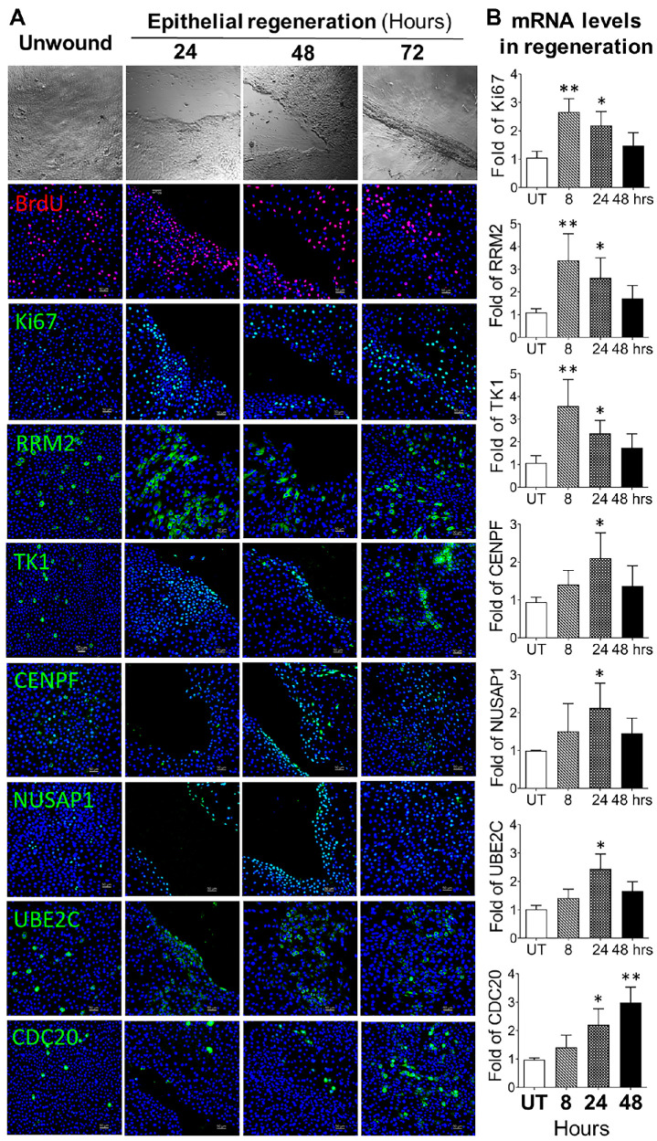
Figure 7.
TAC markers were highly activated during limbal epithelial regeneration in vitro. (A) Representative images show the BrdU-incorporated cells and protein levels of TAC markers during epithelial regeneration after wounding in HLECs. (B) RT-qPCR determined the mRNA expression of Ki67, RRM2, TK1, CENPF, NUSAP1, UBE2C, and CDC20 during the regeneration process (n = 5). Data are shown as mean ± SD. *P < 0.05; **P < 0.01.

