Figure 2.
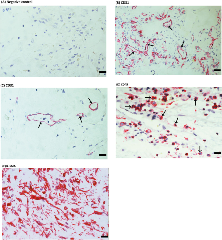
Immunohistochemical staining of proliferative diabetic retinopathy (PDR) epiretinal fibrovascular membranes. (A) Negative control slide (procedure without the addition of the primary antibody) showing no labelling. Immunohistochemical staining for the endothelial cell marker CD31 showing pathologic new blood vessels expressing this endothelial cell marker in a membrane from a patient with active neovascularization (arrows) (B) and in a membrane from a patient with involuted PDR which is composed mostly of fibrous tissue (arrows) (C). Immunohistochemical staining for the leukocyte common antigen CD45 showing infiltrating leukocytes in the stroma (arrows) (D). Immunohistochemical staining for α-smooth muscle actin (α-SMA) showing immunoreactivity in spindle-shaped myofibroblasts (E) (scale bar, 10 µm).

