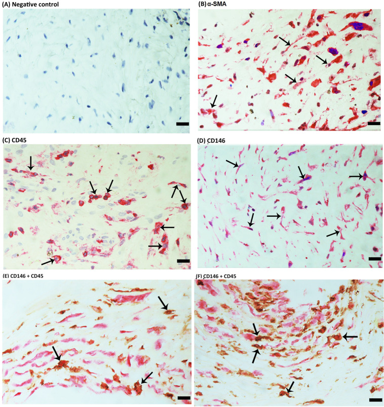
Figure 4.
Immunohistochemical staining of proliferative vitreoretinopathy epiretinal fibrocellular membranes. Negative control slide showing no staining (A). Immunohistochemical staining for α-smooth muscle actin (α-SMA) showing immunoreactivity in myofibroblasts (arrows) (B). Immunohistochemical staining for CD45 showing immunoreactivity in leukocytes (arrows) (C). Immunohistochemical staining for CD146 showing immunoreactivity in spindle-shaped myofibroblasts (arrows) (D). Double immunohistochemistry for CD45 (brown) and CD146 (red) showing cells co-expressing CD45 and CD146. No counterstain to visualize the cell nuclei was applied (arrows) (E, F) (scale bar, 10 µm).

