Figure 5.
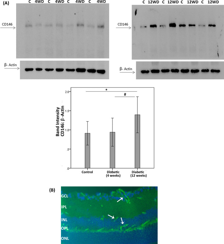
CD146 protein expression in the retinas of diabetic rats. (A) CD146 protein expression was determined by Western blot analysis on lysates of diabetic (D) and nondiabetic control retinas (C) at 4 weeks (4W) and 12 weeks (12W) after diabetes induction. After determination of the intensity of the CD146 protein band, intensities were adjusted to those of β-actin in the sample. Results are expressed as mean ±SD. One-way ANOVA and independent t-tests were used for comparisons between the three and two groups, respectively. *p < 0.05 compared with the values obtained from nondiabetic controls. #p < 0.05 compared with 4 week diabetic rats. (B) Immunofluorescence detection of CD146 (light green) in 8-week diabetic rat retina. CD146 immunoreactivity is detected in endothelial cells of the capillaries (white arrows). Nuclei were counterstained with DAPI (blue). GCL = ganglion cell layer; IPL = inner plexiform layer; INL = inner nuclear layer; OPL = outer plexiform layer; ONL = outer nuclear layer.

