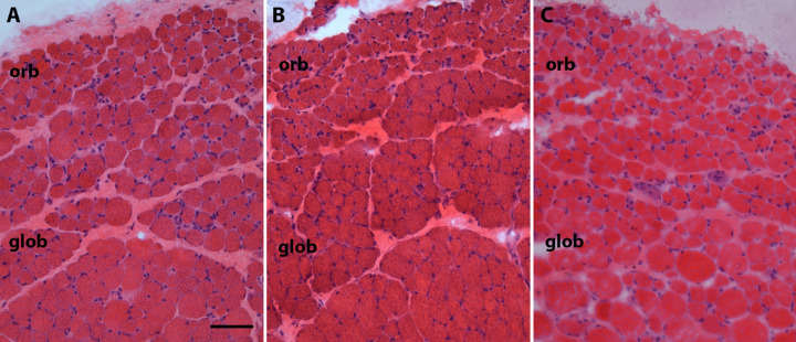
Figure 6.
Photomicrograph of hematoxylin and eosin stained representative sections of superior rectus muscles from (A) naïve control, (B) placebo-pellet treated control, and (C) FGF2-containing sustained release pellets 3 months after treatment. Please note the heterogeneity of the myofibers including very small fibers in C. orb. orbital layer; glob, global layer. Bar is 50 µm.

