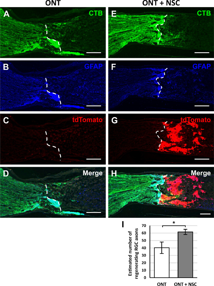
Figure 4.
Optic nerve transplanted NSCs following ONT promote modest, short-distance RGC axon regeneration. Representative images from longitudinal optic nerve sections that received an (A–D) optic nerve transection (ONT) or (E–H) an ONT with NSC graft. Dashed white lines demarcate the areas in which CTB labeling was quantified. (A, E) Intravitreal CTB injections label regenerated RGCs axons at the transection site. (B, F) Immunostaining for GFAP identifies the host tissue and was used to demarcate the proximal border of the transection (white line dotted). (C, D) NSCs constitutively express tdTomato to identify the location and extent of the cell graft. (D, H) Merged images demonstrate the localization of CTB-labeled axons, tdTomato NSCs, and GFAP borders. (I) Quantification of CTB-positive RGC axons beyond the proximal lesion border (dashed line) showed a modest increase in regenerating RGC axons when a NSC graft was present. *P < 0.05; Student's t-test. n = 6 animals per group; error bars represent SD. Scale bars: 200 µm. ONT, optic nerve transection.

