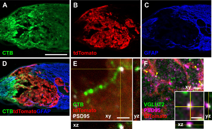
Figure 6.
Optic nerve transplanted NSCs form synapses with transected RGC axons. (A–D) Longitudinal optic nerve section 2 weeks after optic nerve transection and grafting of tdTomato-expressing NSCs in the injury site. (A) Cholera toxin B (CTB) labeled RGC axons regenerate into the injury site. (B) NSCs expressing tdTomato transplanted into the transected optic nerve survive at the transection site. (C) GFAP labeling demarcates the glial response to injury and the boundaries of the optic nerve transection. (D) Merged images demonstrate CTB labeled RGC axons regenerating into the NSC graft. (E) Confocal image of a CTB-labeled RGC axon within the tdTomato-expressing NSC graft in proximity to the postsynaptic marker PSD-95, suggestive of synapses within the NSC graft. (F) Confocal image of VGLUT2 and PSD95 immunostaining within the tdTomato-expressing NSC graft demonstrating overlap of presynaptic and postsynaptic markers (yellow arrowheads); (F inset) high magnification of a puncta of VGLUT2 and PSD95 overlap. Scale bar: 200 µm (A–D); 2 µm (E, F); 1 µm (F inset).

