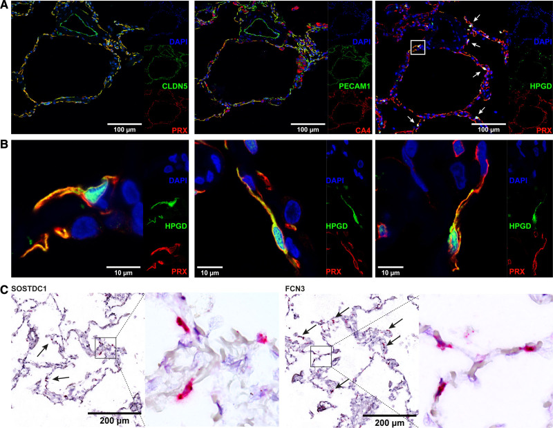
Figure 4.
Localization of aerocytes and general capillary endothelial cells (ECs). A, Representative serial immunofluorescent images of microvascular markers PRX and CA4 with positive red staining in capillaries (CA4 with off-target positive staining in macrophages) and negative staining of a larger central vessel. The general endothelial markers CLDN5 and PECAM1 in green stain the larger central vessels as well, in addition to the microvasculature. The third image shows an immunostain of the aerocyte-specific marker HPGD in green (white arrows), in addition to general microvascular marker PRX in red. Nuclei are counterstained throughout with DAPI (4′,6-diamidino-2-phenylindole). The white box highlights an aerocyte, which is shown at higher magnification in the first image of B. B, Representative immunostains of aerocytes with the specific marker HPGD in green (cytoplasmic and nuclear expression pattern), which colocalizes with the general microvascular marker PRX in red. Nuclei are counterstained throughout with DAPI. C, In situ RNA hybridization stains of markers specific to the capillary subpopulations with SOSTDC1 staining aerocyte ECs (arrows) and FCN3 staining general capillary ECs (arrows) with positive staining in red. The black box highlights an area shown to the right of it in high magnification. Conventional immunohistochemical images of shown markers can be found in Figure IV in the Data Supplement.

