Figure 5.
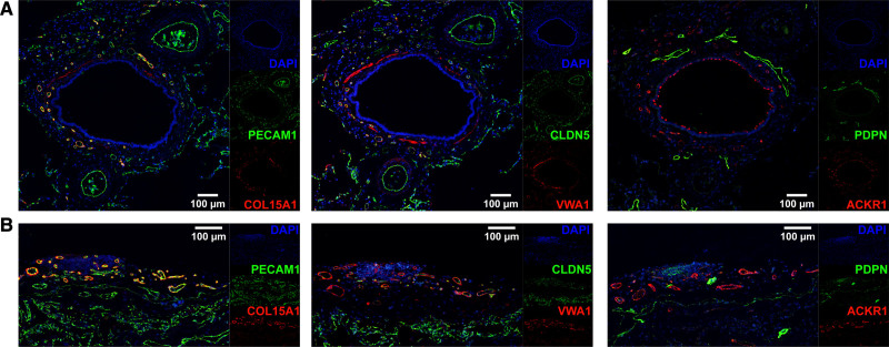
Systemic–venous endothelial cells (ECs) localize to the bronchial vascular plexus and visceral pleura. Representative, serial immunofluorescent images of systemic–venous EC markers COL15A1 and VWA1, panvenous marker ACKR1 (all in red), panendothelial markers PECAM1 and CLDN5, and lymphatic marker PDPN (all in green) in (A) a bronchus with accompanying arteries and (B) visceral pleura. In (A), COL15A1, VWA1, and ACKR1 stain the small vessels of the bronchial vascular plexus, but not the accompanying arteries; PECAM1 and CLDN5 stain all vessels. In (B), COL15A1, VWA1, and ACKR1 stain the pleural vessels, but not the alveolar microvasculature; PECAM1 and CLDN5 stain all vessels. PDPN identifies bronchial (A) and pleural (B) lymphatic vessels. VWA1 exhibits off-target staining in smooth muscle cells, and ACKR1 in ciliated cells. Nuclei are counterstained throughout with DAPI (4′,6-diamidino-2-phenylindole). Conventional immunohistochemical images of shown markers can be found in Figure IV in the Data Supplement.

