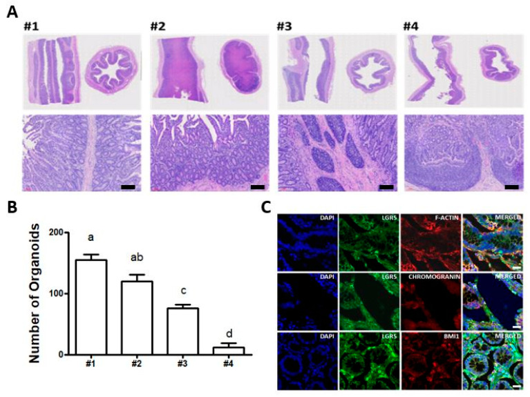
Figure 2.
Immunohistochemical analysis of bovine small intestine: (A) haematoxylin and eosin histological staining to identify distinct crypt and villus structures from four different locations (#1, #2, #3, and #4) in the jejunum between the duodenum and ileum. Scale bar: 300 μm; (B) the number of intestinal organoids per basement matrix dome to verify the efficiency of the derivation of intestinal organoids from four different locations. The number of intestinal organoids derived from location #1 in the jejunum close to the duodenum was significantly higher, compared to locations #3 and #4. The values are the means plus the standard error of mean (S.E.M) and different letters (a–d) indicate significant differences (p < 0.05); (C) immunohistochemistry of LGR5, Bmi1, F-actin, and Chromogranin A in bovine small intestine. The fluorescently stained crypts were counterstained with diamidino-2-phenylindole (DAPI). Scale bar: 20 μm.

