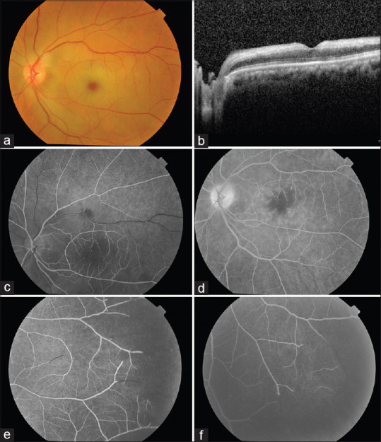
Figure 1.

(a) Fundus photograph of the left eye shows a cherry-red spot at the macula. Note the unusual supply to the superior quadrant from the inferotemporal branch retinal artery; (b) Optical coherence tomography macula showing retinal inner layer hyperreflectivity due to retinal edema; (c) Early phase of fundus fluorescein angiogram showing enlarged foveal avascular zone suggestive of ischemic macula; (d) Late phase showing diffuse leakage due to retinal edema. Note the incomplete terminal filling of the branch arteries supplying the macula; (e and f) Fundus fluorescein angiogram of the peripheral retina showing incomplete arterial filling with large capillary nonperfusion areas
