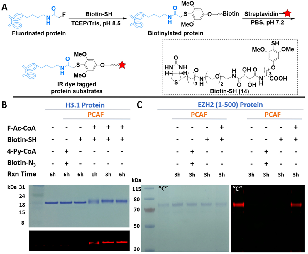
Figure 3.

FTDR-based tagging of protein substrates with biotin–SH probe. (A) Reaction scheme. The red star indicates IR dye. (B, C) Labeling of histone protein H3.1 and nonhistone substrate EZH2 (1 – 500), respectively. The top panel for (B) and the left panel for (C) are gel images after CBB staining. The other images are for in-gel fluorescent detection of IR dye. “C” is the positive control for CuAAC, which has been prepared by NHS ester labeling of the lysines on EZH2 with the “click chemistry” tag, followed by CuAAC reaction with the corresponding biotin linker.
