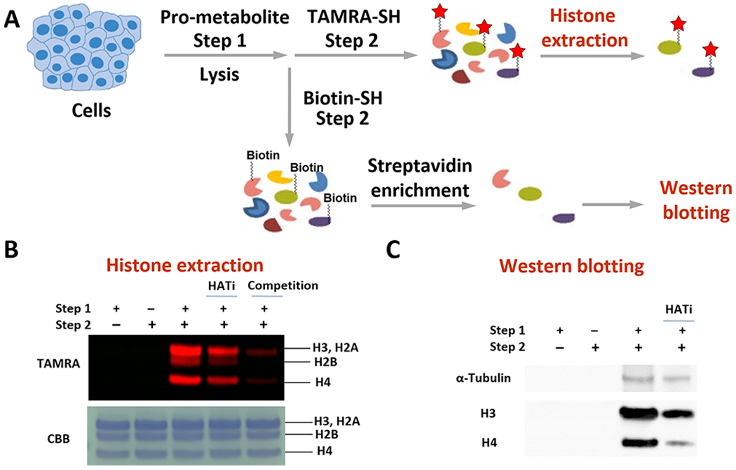
Figure 5.

Validation of the FTDR-based labeling of acetylation substrates. (A) Scheme for cellular pro-metabolite incorporation (step 1), protein substrate labeling by TAMRA–SH (step 2), and extraction of the known acetylation substrates histones; or protein substrate labeling by biotin–SH probe (step 2), enrichment with streptavidin beads, followed by Western blot analysis of the proteins pulled down to examine the existence of α-tubulin, histone H3, and histone H4. (B) Histone extraction results. Top panel, in-gel fluorescent detection; bottom panel, CBB. (C) Western blotting results. “HATi” indicates the addition of anacardic acid and MG149.50–52
