Figure 2.
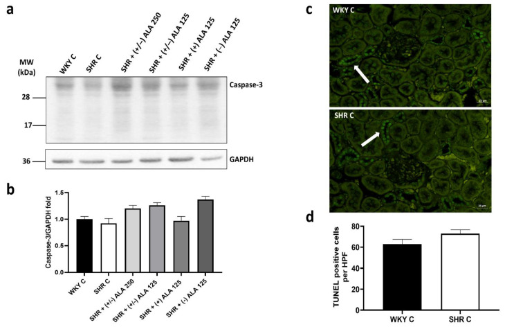
Apoptosis in the kidney. (a) Lysates of kidney from WKY C, SHR C, SHR + (+/−) alpha-lipoic acid (ALA) 250 µmol/kg/day, SHR + (+/−) ALA 125 µmol/kg/day, SHR + (+) ALA 125 µmol/kg/day, SHR + (−) ALA 125 µmol/kg/day were immunoblotted using specific anti caspase-3; (b) The bar graph indicates the ratio of densitometric analysis of bands and GAPDH levels, used as loading control, considering the WKY C group as reference. Blots are representative of one of three separate experiments; (c) Sections of kidney from WKY C and SHR C processed for TUNEL staining. Arrow: apoptotic nucleus. Calibration bar 25 µm; (d) Bar graph shows the quantification of the number of positive cells per high-power field (HPF) (40×). Data are mean ± S.E.M.

