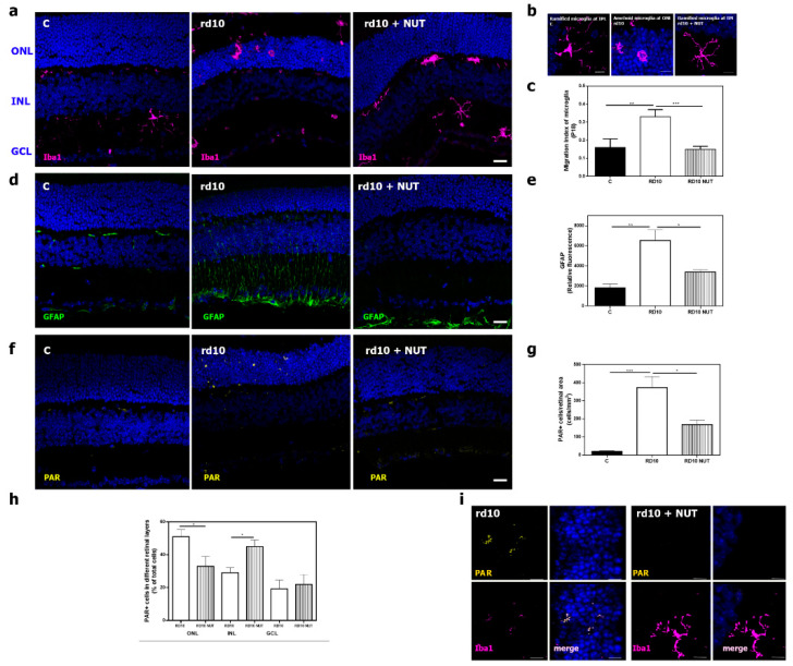
Figure 4.
Effect of oral administration of nutraceuticals on retinal inflammation in rd10 mice at PD18: (a) representative photomicrographs of retinal sections showing Iba1-labeling (microglial cells) in DAPI-counterstained section from control mice (C), untreated rd10 mice, and NUT-treated rd10 mice. rd10 were treated with NUT from PD9 to PD18, scale: 20 µm; (b) optical zoom showing amoeboid and ramified shape of microglia cells in retinas of untreated rd10 mice and NUT-treated rd10 mice, scale: 10 µm; (c) index of microglial migration in these retinas; (d,e) representative photomicrographs of retinal sections showing GFAP-labeling (Müller cells) in DAPI-counterstained sections and corrected fluorescence from control (C), untreated rd10 and NUT-treated rd10 mice, scale: 20 µm; (f,g) representative photomicrographs of retinal sections showing poly ADP-ribose polymers (PAR) accumulation, quantification of PAR positive cells from control (C), untreated rd10 and NUT-treated rd10 mice scale: 20 µm; (h) quantification of the distribution of PAR positive cells throughout the retina of untreated rd10 and NUT-treated rd10 mice, (i) optical zoom showing double-immunostaining of Iba1 and PAR in retinas of untreated rd10 mice and NUT-treated rd10 mice, scale: 5 µm. ONL: outer nuclear layer; INL: inner nuclear layer; GCL: ganglion cell layer. Data were presented as mean ± SEM. from at least eight retinas for each experimental group. Statistical differences between groups (p < 0.05) were shown * p < 0.05; ** p < 0.01; *** p < 0.001 using one-way ANOVA or Kruskal–Wallis test and post hoc Tukey’s or Dunn’s multiple comparisons tests.

