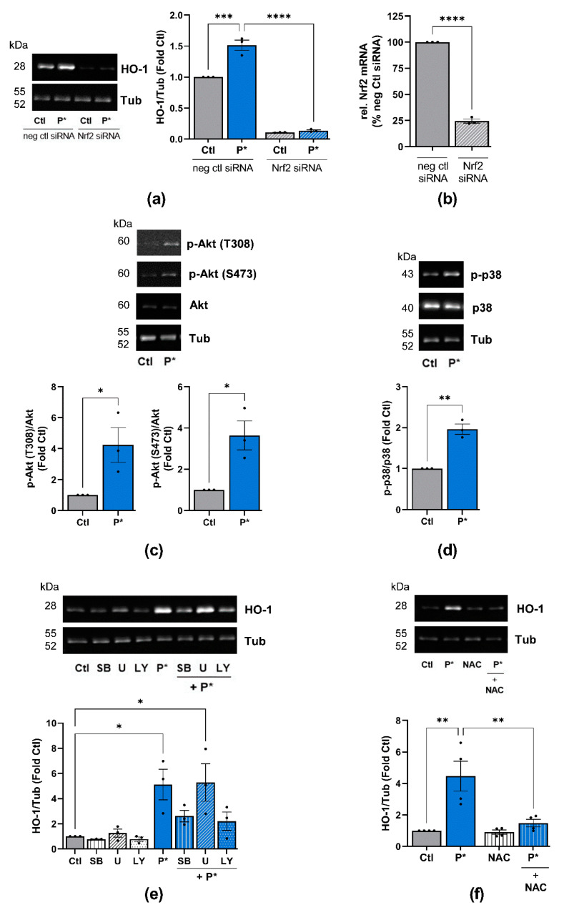
Figure 5.
P*-mediated HO-1 induction requires activation of Nrf2, PI3K/Akt, and p38 MAPK pathways. (a) ATDC5 cells transfected with Nrf2 (Nrf2 siRNA) or negative control (neg ctl siRNA) siRNA were preincubated with 0.5 mM P* for 6 h. Expression of HO-1 was analyzed by Western blotting using α, β-tubulin (Tub) as control. Representative Western blots are shown. Densitometry analysis values of HO-1 were normalized against Tub and expressed as relative quantity compared to untreated cells (Ctl) set to 1, n = 3, *** p < 0.001, **** p < 0.0001. (b) Knockdown efficiency of Nrf2 by siRNA was validated by qPCR with cyclophilin D as a reference gene, n = 3, **** p < 0.0001. (c,d) ATDC5 cells were incubated with 0.5 mM P* for 6 h, and protein expression was analyzed through Western blotting. Representative Western blots of the expression of (c) p-Akt, Akt and Tub or (d) p-p38, p38 MAPK and Tub are shown. Densitometry analysis values of phosphorylated proteins were normalized against equivalent unphosphorylated proteins and then compared to untreated cells (Ctl) set to 1, n = 3, * p < 0.05, ** p < 0.01 (unpaired t-test). (e,f) Representative Western blots of the expression of HO-1 and Tub. Densitometry analysis values of HO-1 were normalized against Tub and expressed as relative quantities compared to Ctl set to 1, n = 3, * p < 0.05, ** p < 0.01. ATDC5 cells were incubated for 1 h with (e) p38 MAPK inhibitor SB203580 (SB, 10 µM), MEK1/2 inhibitor U0126 (U, 10 µM), or PI3K inhibitor LY294002 (LY, 10 µM), or (f) 2 mM N-acetylcysteine (NAC), followed by 6 h incubation with 0.5 mM P*.

