
FIGURE 2.
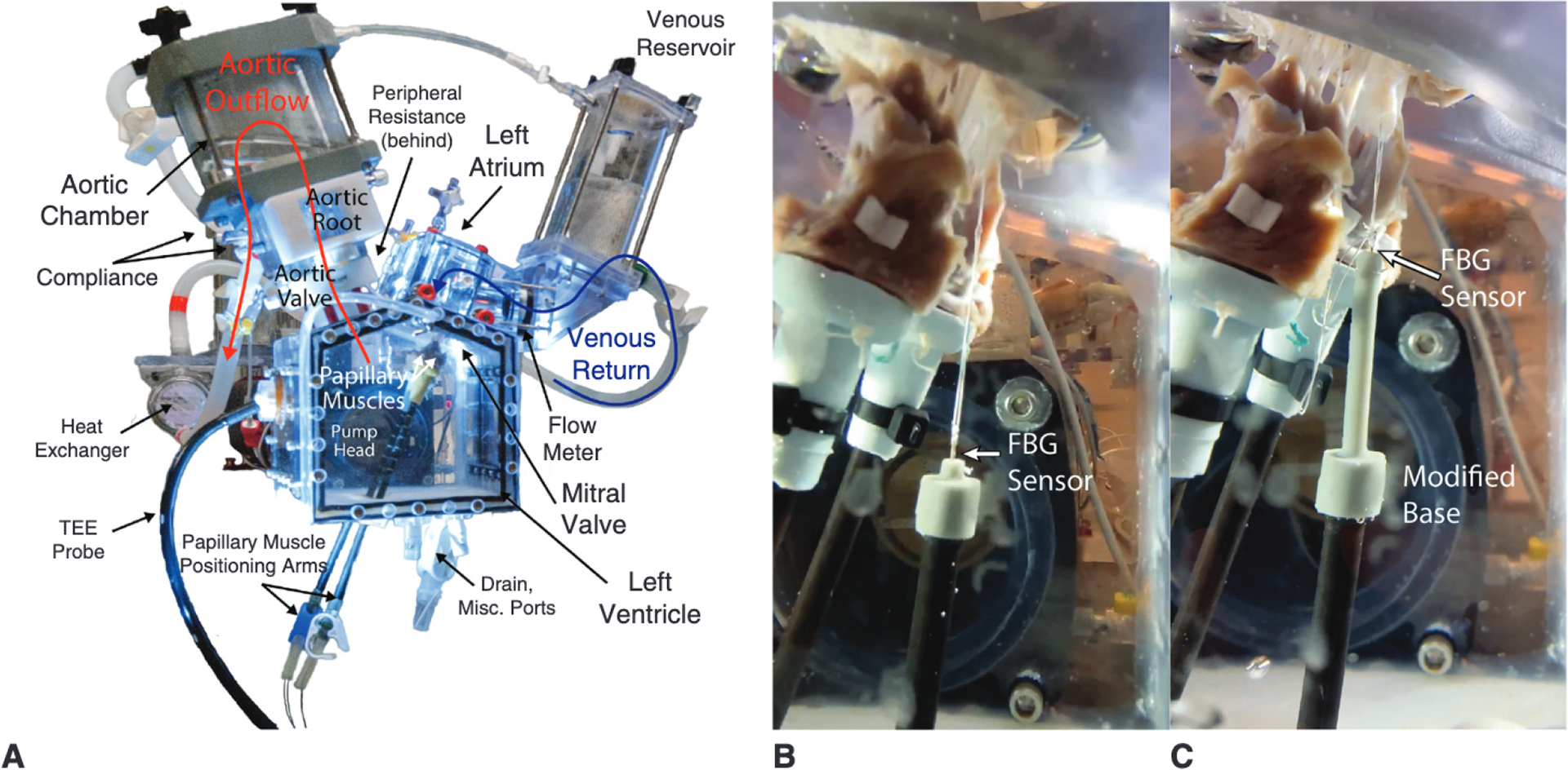
A, Labeled image of the heart simulator featuring compliance chambers and adjustable resistive elements, and including a left ventricular chamber affixed to a linear piston pump programmed to reproduce physiologic pressures and flows. B, Heart simulator testing of a mitral valve repaired with a Gore-Tex CV-5 suture anchored to a custom force-sensing post positioned to mimic apical placement. C, Without adjusting the post position, the same valve was tested with the artificial papillary muscle device serving as the suture anchor. A low-profile and high-resolution FBG sensor was used in each case to measure real-time artificial chordae forces. TEE, Transesophageal echocardiography; FBG, fiber bragg grating.