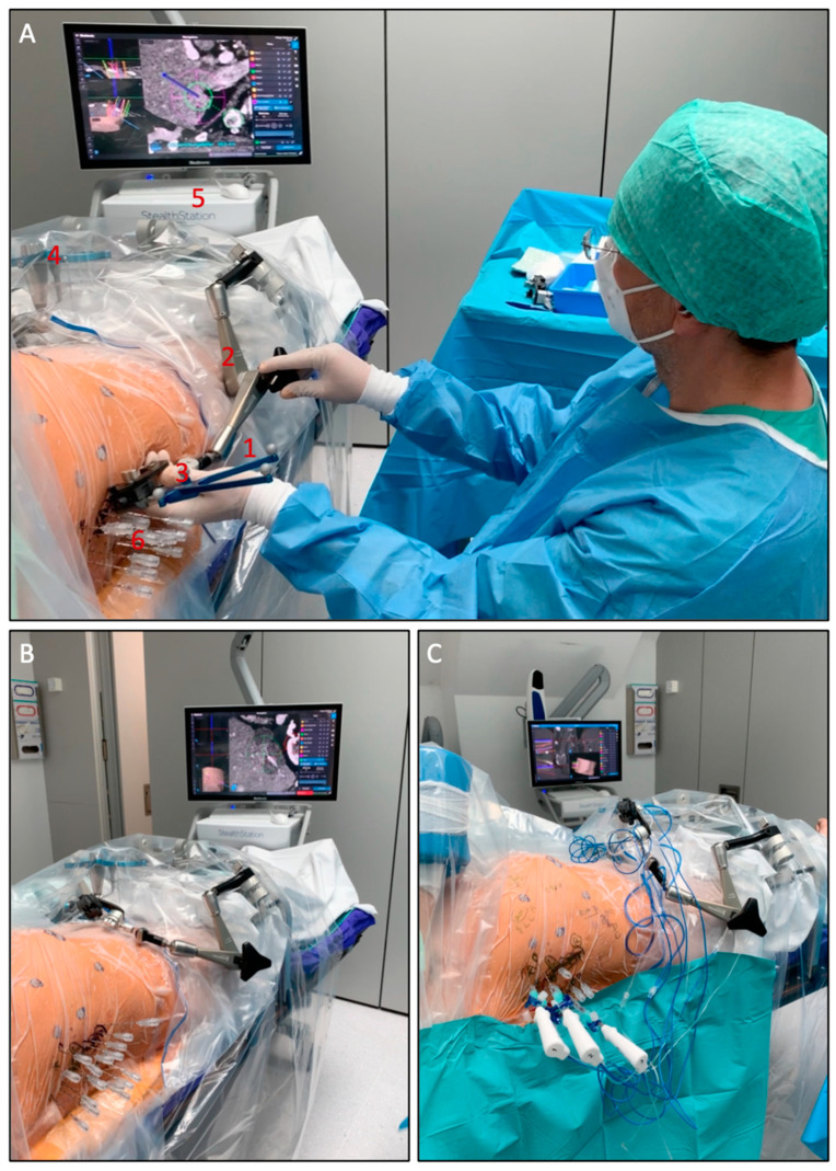
Figure 1.
Setup of the SRFA procedure. (A) The probe (1) of the navigation system is inserted into the ATLAS aiming device (2). The reflective markers (3) on the probe and the reference frame (4) are tracked by the camera of the navigation system (5). After manual alignment of the aiming device with the virtual trajectory and consecutive removal of the probe, the interventionalist introduces several coaxial needles (6) through the locked aiming device to the preplanned target point. (B) Placed coaxial needles in final position. (C) After verification of correct needle placement with a non-enhanced control CT scan and image fusion with the planning-CT up to three RF-electrodes at a time are introduced through the coaxial needles for serial tumor ablation.

