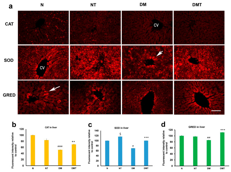
Figure 3.
Fluorescence images (a) of catalase (CAT), superoxide dismutase (SOD), and glutathione reductase (GRED) expression in the liver of normal (N), normal treated (NT), diabetic untreated (DM), and diabetic treated with nociceptin (DMT). Note that the expression of SOD was significantly higher in normal and diabetic rats treated with nociceptin. Nociceptin also markedly increased the tissue level of GRED after nociceptin treatment in diabetic rats (b–d). cv = central vein; n = 6; Scale bar = 25 µm; Magnification = ×400; $ (normal treated versus normal untreated); ## and ### (diabetic untreated versus normal untreated); ** and *** (diabetic untreated versus diabetic treated). $ p < 0.05; # p < 0.05; ## p < 0.01; ### p < 0.001; ** p < 0.05; *** p < 0.001.

