Figure 8.
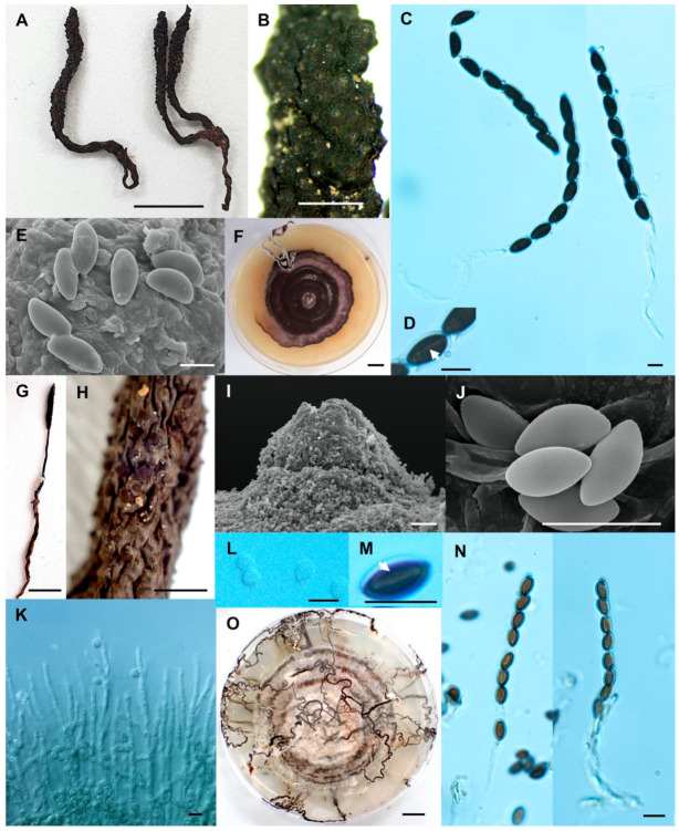
Xylaria sihanonthii (SWUF18-1.3). (A) Stromata. (B) Stromatal surface with ostioles. (C) Asci with ascospores. (D) germ slits (arrowed). (E) Ascospores. (F) Colony on PDA in a 9 cm Petri dish at 4 weeks. Xylaria subintraflava (PK17-24.2). (G) Stroma. (H) Wrinkled stromata surface with ostioles. (I) Ostiole. (J) Ascospores. (K) Anamorph. (L) Conidia. (M) Germ slit (arrowed). (N) Asci and ascospores. (O) Colony on PDA in a 9 cm Petri dish at 4 weeks. (E,I,J) by SEM; (C,D,K–N) by DIC. Scale bars (A,F,G,O) = 1 cm; (B,H) = 1 mm; (C–E,J–N) = 5 µm; (I) = 20 µm.

