Figure 6.
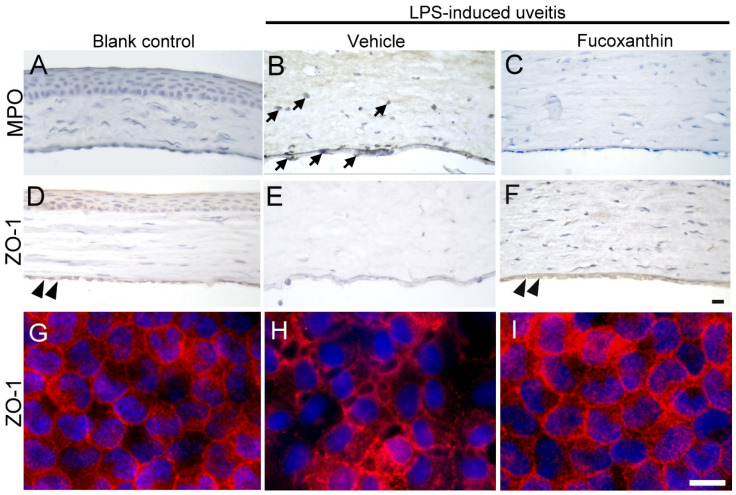
Effects of fucoxanthin on the infiltration of LPS-induced MPO-positive cells and disruption of ZO-1 in the corneal endothelium of the anterior chamber. MPO (A–C) and ZO-1 (D–I) expression levels among the blank control (A,D,G), LPS/vehicle (B,E,H), and LPS/10 mg/kg fucoxanthin (C,F,I) groups. Immunohistochemical staining showed strong adherent effects and infiltration of MPO-positive inflammatory cells (arrows) in the LPS/vehicle group (B) relative to equivalent measurements in the control group (A). In contrast, there were decreased numbers of MPO-positive inflammatory cells in the anterior chamber in the fucoxanthin/LPS group (C). Moreover, histological examination showed a continuous brown stained ZO-1 in the region of cell–cell contact in the corneal endothelium of the blank control (double arrowheads, (D)). Disruption of ZO-1 was observed in the LPS/vehicle group (E) relative to in the control group. Compared to the LPS group (E), an increase in the intensity of browning staining ZO-1 expression at the cell–cell junction was noted in the LPS/fucoxanthin group (double arrowheads, (F)). On whole flat-mounted corneas, disruption of tight junction was prevented in the LPS/10 mg/kg fucoxanthin group (G–I). Nuclei were stained with hematoxylin or DAPI. Scale bars = 20 μm.

