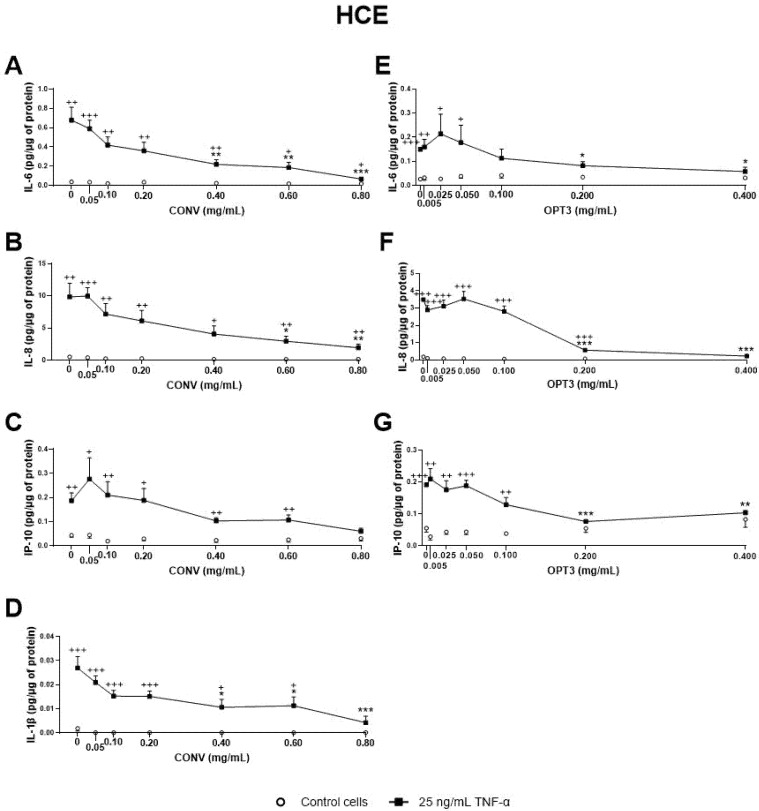
Figure 3.
Effect of the conventional (CONV) and the selected optimized (OPT3) olive pomace (OP) extracts on TNF-α-induced cytokine release by HCE cells. Cells were pre-treated with CONV (0.05, 0.10, 0.20, 0.40, 0.60, and 0.80 mg/mL), OPT3 (0.005, 0.025, 0.050, 0.100, 0.200, and 0.400 mg/mL), or vehicle (0.4% ethanol) for 2 h. Following this, they were stimulated with 25 ng/mL TNF-α in the presence of the treatments for 24 h (black squares). Vehicle-treated-TNF-α stimulated cells and cells not stimulated with TNF-α (white circles) were used as control. IL-6, IL-8, IP-10, and IL-1β were measured in cell supernatants by a multiplex bead-based array. TNF-α failed to stimulate IL-1β in the experiment performed for OPT3. CONV significantly decreased IL-6 levels from 0.40 mg/mL (A), IL-8 levels from 0.60 mg/mL (B), and IL-1β levels from 0.40 mg/mL (D). For TNF- α stimulated cells, IP-10 production was not decreased significantly by CONV. Nevertheless, no significant differences were observed between stimulated and non-stimulated cells at 0.80 mg/mL (C). OPT3 inhibited IL-6 (E), IL-8 (F), and IP-10 (G) secretion from 0.200 mg/mL significantly. Data are presented as picograms (pg) of cytokine/chemokine per micrograms (μg) of total protein for three independent experiments (performed in duplicate) ± SEM. * p < 0.05, ** p < 0.01, *** p < 0.001, compared with vehicle-treated-TNF-α stimulated cells; + p < 0.05, ++ p < 0.01, +++ p < 0.001, compared with control cells.

