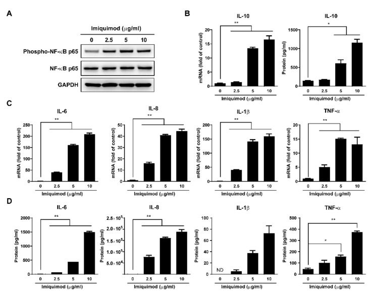
Figure 6.
Effect of imiquimod on NF-κB signaling pathway. (A) Macrophages were treated with imiquimod at indicated concentrations for 24 h. Then cellular protein was collected and subjected to Western blot for GAPDH, NF-κB p65, and Phospho-NF-κB p65 expression. (B) Macrophages were treated with imiquimod at indicated doses for 6 h or 24 h, and then cellular RNA or culture supernatant was collected and subjected to real-time PCR or ELISA for IL-10 expression. (C) Macrophages were treated with imiquimod at indicated concentrations for 6 h. Then cellular RNA was collected and subjected to real-time PCR for IL-6, IL-8, IL-1β, and TNF-α expression. (D) Macrophages were treated with imiquimod at indicated concentrations for 24 h. Then culture supernatant was collected and subjected to ELISA for IL-6, IL-8, IL-1β, and TNF-α expression. Data are shown as mean ± SD of three independent experiments (ND: not detected, * p < 0.05, ** p < 0.01).

