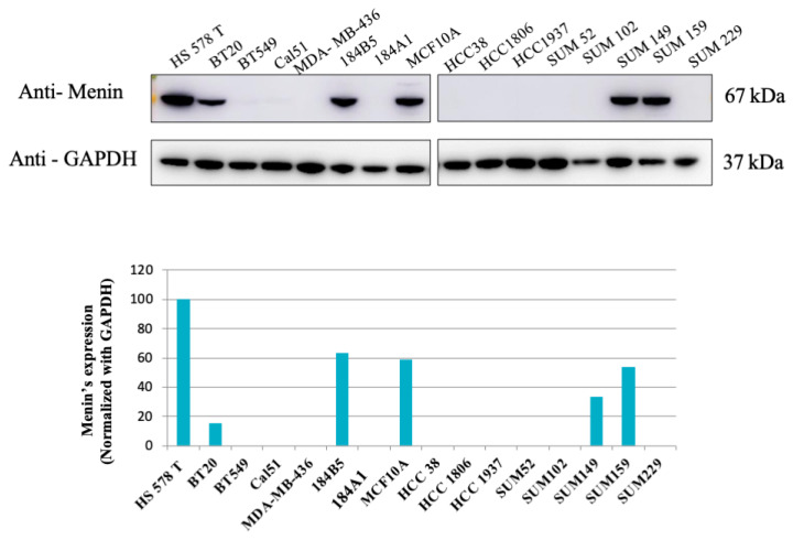
Figure 1.
Western blot analysis of Menin expression in 16 TNBC cell lines. The expression of menin in 16 TNBC (triple-negative breast cancer) cell lines were evaluated using WB analyses. Six out of sixteen cell lines displayed menin’s expression, and Hs 578T had the highest level of menin. Bands were quantified by densitometry and menin was normalized to GAPDH levels.

