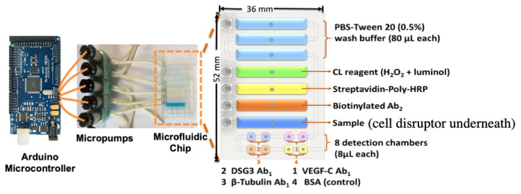
Figure 7.
Microfluidic immunoarray for cell disruption and protein detection. The design features a microfluidic chip with five inlets connected to peristaltic micropumps, sample and rectangular prism reagent chambers with capacity of 80 ± 5 μL, and eight cylindrical detection chambers with 8 ± 1 μL capacity each. The microfluidic chip houses sample and reagents and delivers them sequentially to the detection compartment. The assay protocol utilizes poly-HRP and ultra-bright femto-luminol to produce chemiluminescence (CL) that is captured in a dark box using a CCD camera. The microfluidic chip is mounted on the housing device support equipped with a sonic cell disruptor that achieves cell lysis. Programmable micropumps are connected to microfluidic chip sample and reagent chambers and the assay is automated by using an Arduino microcontroller that control pump on/off cycles. Adapted from [43], with permission. Copyright Elsevier, 2021.

