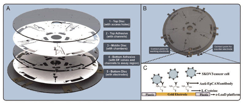
Figure 10.
(A) Rendered 3D image of the five-layer microfluidic disc platform comprising three 1.5-mm thick PMMA discs and two 90-μm thick pressure sensitive adhesive films. The gold electrodes were deposited on the bottom (layer 5) of the disc. (B) Fully assembled disc showing contact points for the working and counter electrodes. (C) Schematic of electrochemical cancer cell capture assay on polymeric eLoaD platform. Reproduced with permission from [76].

