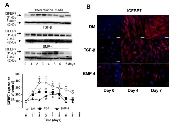
Figure 3.
IGFBP7 expression is increased in human ASCs going through differentiation with TGF-β and BMP-4. (A) Top is representative immunoblot for IGFBP7 expression. Corresponding line graph demonstrating the time-course (0 to 7 days) effect of TGF-β and BMP-4 on IGFBP7 expression. TGF-β and BMP-4 synergistically induce the expression of IGFBP7 on hASC. hASCs were stimulated with TGF-β, BMP-4 or combination of TGF-β and BMP-4 (DM) for up to 7 days and IGFBP7 expression was evaluated by Western blot. Top are representative immunoblots for IGFBP7 expression. Corresponding bar graph demonstrating the time-course (0 to 7 days) effect of TGF-β, BMP-4 or DM on IGFBP7 expression. (B) Fluorescence microscopy was also used to detect TGF-β, BMP-4 or DM-induced IGFBP7 expression on day 0, 4 and 7. Scale bar 100µm. Results are mean ± SEM of 5 experiments. * p < 0.05 vs. day 0 of stimulation with DM; ** p < 0.05 vs. day 0 of stimulation with TGF-β; # p < 0.05 vs. day 0 of stimulation with BMP-4.

